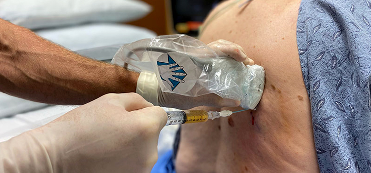
Pleural Fluid Tapping
A thoracentesis (pleural tap) is a procedure that removes fluid from around the lungs, or pleural fluid. Your doctor may recommend a thoracentesis to diagnose and guide treatment for certain diseases, such as cancer, infections, and heart failure.
Pleural fluid cushions and lubricates the lungs as they expand and contract with breathing. Pleural fluid is located in the pleural space, the space inside the lining that covers the lungs and inside of your chest. Certain diseases and conditions cause a buildup of pus, blood, or too much pleural fluid in the pleural space. This is called pleural effusion, which can put pressure on the lungs and make it difficult to breathe.
A thoracentesis is only one method of diagnosing and treating pleural effusion and underlying problems. Discuss all of your testing options with your doctor to understand which options are right for you.
Why is a thoracentesis (pleural tap) performed?
Your doctor may recommend a thoracentesis (pleural tap) to diagnose or treat pleural effusion and empyema (pronounced “em-pie-eema”). Pleural effusion is a buildup of fluid in the pleural space, inside the lining that covers the lungs and inside of the chest. Empyema is pus in the pleural space.
A thoracentesis removes fluid to make it easier for you to breathe. Your doctor may also test the fluid to determine the cause of pleural effusion or empyema. A thoracentesis helps your doctor make the best treatment decisions for underlying diseases, disorders and conditions including:
- Heart failure, an inability of a weak heart to pump enough blood to the body. This can lead to fluid buildup.
- Pulmonary hypertension, high pressure in the arteries that carry blood to the lungs. Pulmonary hypertension can lead to heart failure and fluid buildup.
- Pulmonary embolism, a blood clot or other blockage in the artery that carries blood to the lungs. Pulmonary embolism can lead to heart failure, pulmonary hypertension, and fluid buildup.
- Nephrotic syndrome, kidney damage that causes fluid buildup
- Lung infections including pneumonia and pleurisy, which can cause inflammation of the pleural lining, fluid buildup, and sometimes pus
- Cancer that has spread to the lung or the pleural lining. The pleural fluid is tested for cancer cells.
- Liver disease including liver cirrhosis and liver failure, which can cause fluid buildup
How is a thoracentesis (pleural tap) performed?
Your thoracentesis will be performed in a hospital, clinic, or doctor’s office. The procedure generally takes 15 minutes and includes these steps:
- You will remove your clothing and dress in a patient gown.
- You will have an imaging test, such as an X-ray, ultrasound, or CT scan of the chest to locate the fluid and see how much fluid is around the lungs.
- You will sit on a chair or edge of an examination table with your arms resting on a table. You must remain very still during the entire procedure. Do not cough or breathe very deeply.
- Your doctor will clean the procedure area on your back with an antiseptic and place sterile drapes on your back to maintain a sterile procedure.
- Your doctor will inject a small amount of local anesthetic under the skin to numb the area. In some cases, you may receive a sedative to help you relax.
- Your doctor will insert a needle or tube into your back between the ribs and into the pleural space around the lungs. Sometimes doctors use ultrasound imaging to help place the needle in the right place.
- Your doctor will remove pleural fluid for testing and drain excessive pleural fluid as needed.
- Your doctor will remove the needle, bandage the area, and send the fluid to the laboratory for testing.
- Your care team will watch your vital signs closely for several hours after your thoracentesis. You will have a chest X-ray to look for complications, such as a collapsed lung (pneumothorax).
What are the risks and potential complications of a thoracentesis (pleural tap)?
Complications of a thoracentesis are uncommon, but any procedure involves risks and potential complications that may become serious in some cases. Complications can develop during the procedure or your recovery.
Risks and potential complications of a thoracentesis include:
- Adverse reaction or problems related to sedative or anesthetic medications such as an allergic reaction and problems with breathing
- Bleeding
- Collapsed lung (pneumothorax)
- Infection
- Injury to the liver or spleen, which is very rare
Reducing your risk of complications
You can reduce the risk of certain complications by following your treatment plan and:
- Following activity, dietary and lifestyle restrictions and recommendations before your procedure and during recovery
- Informing your doctor or radiologist if you are nursing or if there is any possibility of pregnancy
- Notifying your doctor immediately of any concerns, such as difficulty breathing or pain or discomfort that does not pass quickly
- Remaining very still and not coughing or taking deep breaths during the procedure as instructed
- Taking your medications exactly as directed
- Telling all members of your care team if you have any allergies
