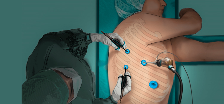
A thoracoscopy is a common procedure to look at the surface of your lungs and the area around your lungs (pleural space). Your healthcare provider uses a thoracoscope (a thin camera with a light) to see these areas and take samples of lung tissue or lymph nodes. They can see your diaphragm, esophagus, chest wall and other areas as well.
Providers use thoracoscopy as part of video-assisted thoracoscopic surgery (VATS), a minimally invasive chest surgery. They can project images to a video monitor in the operating room while they work.
When your provider uses a thoracoscopy to look at your lungs and the areas around them, that’s a medical procedure. They may call it a pleuroscopy to be clear about what they plan to do.
Thoracoscopy is diagnostic when it’s used to look in your chest or take samples (biopsies) of tissue.
Therapeutic thoracoscopy is used as part of minimally invasive surgery to treat a specific problem.
Your healthcare provider can use a thoracoscopy or video-assisted thoracoscopy surgery when they need to:
If you have lung cancer or mesothelioma, you may need this procedure. Also, your provider may use a thoracoscopy when treating cancer in your thymus gland or esophagus.
Thoracoscopy complications may include: