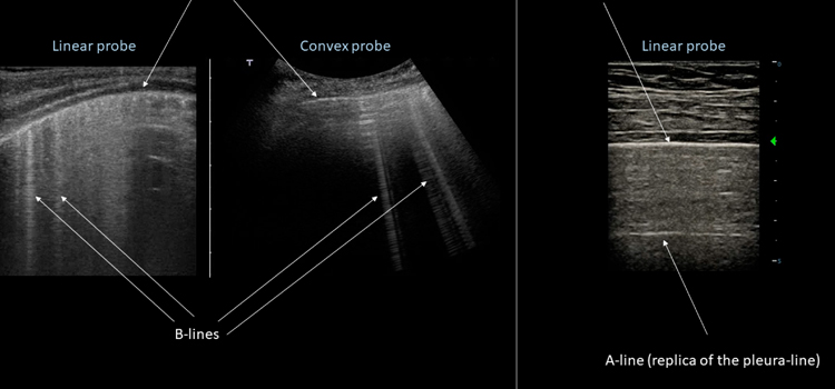
Ultrasound of the thorax is a medical imaging technique that uses high-frequency sound waves to create images of the chest area. It is a non-invasive and safe diagnostic tool that can help evaluate the structures and organs within the chest cavity, such as the lungs, heart, blood vessels, and pleura.
During an ultrasound thorax exam, a small handheld device called a transducer is placed on the patient's chest, which emits high-frequency sound waves that penetrate the body and bounce off the organs and structures within the chest. The sound waves are then detected by the transducer and converted into images by a computer.
Ultrasound thorax can be used to assess various conditions such as pleural effusion, pneumothorax, pulmonary embolism, and pneumonia. It is also useful for monitoring the progression of certain diseases, such as lung cancer and pulmonary hypertension.
Overall, ultrasound thorax is a valuable diagnostic tool that can provide important information about the structures and organs within the chest cavity, and can be used to guide further diagnostic and treatment decisions.
Ultrasound thorax is done for a variety of reasons, depending on the symptoms or conditions being evaluated. Some common reasons for performing an ultrasound of the thorax include:
Overall, an ultrasound thorax is a valuable diagnostic tool that can provide important information to help diagnose and manage a variety of conditions related to the chest and lungs.
Ultrasound thorax is generally considered to be a safe and non-invasive diagnostic procedure with very low risk of complications. Unlike other imaging techniques such as X-rays and CT scans, it does not use ionizing radiation.
There are a few rare and minor risks associated with ultrasound thorax. These may include:
It is important to note that ultrasound thorax is generally considered to be safe for use in pregnant women, as it does not use ionizing radiation. However, as with any medical procedure, it is important to discuss the risks and benefits of the procedure with your healthcare provider before undergoing an ultrasound thorax.